Монгол Xэл (Thanks to Oyu Choijamts for the translation!)
I’ve been out of blogging-commission the past month or so due to a hectic but very productive trip back to the University of Colorado to do some preliminary analysis of some particulate matter samples from Ulaanbaatar and to pick up some lovely new equipment that I was able to get through my new grant. But now that I’m back in UB and the dust has settled from my trip, I wanted to briefly describe some of the cool stuff we’re doing!
In Maggie Tolbert’s lab in CIRES at CU, one of the ways in which we analyzed some Ulaanbaatar particulate matter samples was through Raman spectroscopy on a particle-by-particle basis. What does this mean and why is it so cool? Here’s a short explanation:
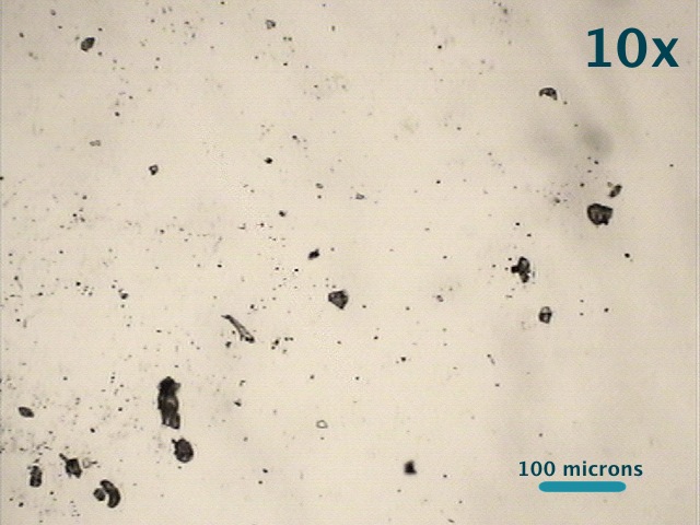
By using a regular microscope, we can look at particulate matter that is on the order of microns or tens of microns in size.
A typical image might look like this:

An example of particulate matter collected in Ulaanbaatar, Mongolia in March 2012. 10x magnification.
Then we can zoom in even closer on one particle:
And the cool part is that we can shoot that particle with a laser, and based on the Raman scattering of the laser light off of the particle, we can get a better idea of the chemical composition of the particle.
For instance, when we shoot the particle above, we get the following Raman spectrum:

The Raman spectrum of the particle shown in the previous image. According to the spectrum, the particle contains organics and carbonates (among other things).
In this spectrum, there’s quite a bit going on, but two notable items are evidence of organics (C-H vibrations – highlighted in blue) and carbonates (highlighted in purple).
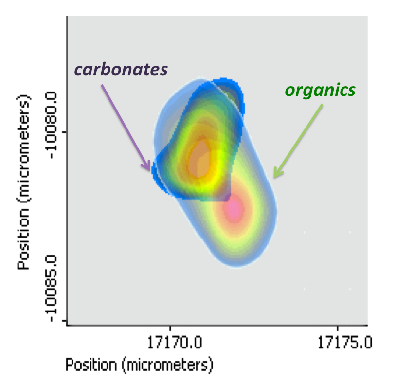
But there’s an even cooler part: We can systematically shoot the particle in various spots and make a 2-D “map” of the particle. If we just focus on the wavenumber regions that are signatures of the organics (remember, the part in blue) and the carbonates (the part in purple), we get a picture like this:

2D Raman map of the 5 micron particle above showing where the carbonate and organic signatures of the particle are located. The warmer colors indicate higher amounts of a particular chemical substance at a given location.
So now we know not only something about the chemical composition of the particle, but also where those constituent components are located within the particle. Knowing the chemical composition of individual particles (or in other words, on a particle-by-particle basis) – and how they are dispersed within the particle itself can indicate many things. It might give a clearer indication of how the particle formed, how it will interact with incoming solar radiation (a particle’s direct effect), and how it might affect the formation of clouds and consequently precipitation (a particle’s indirect effect – to be discussed in more detail in a later post). And of course, this can’t be done for just one particle in a given sample, but many particles chosen at random in order to get meaningful statistics.
At any rate, that is a snippet of what I’ve been up to the past few weeks!
Монгол Xэл:
Сүүлийн сард би Колорадогийн Их Сургуульдаа очиж Улаанбаатараас цуглуулсан PM-ийн (РМ=микро бүрэлдхүүн) сорьцоо анализанд өгөнгөө тэтгэлэгийнхээ тусламжаар авах боломжтой болсон шинэ тоног төхөөрөмжөө аваад УБ-т буцаад ирлээ. Үнэхээр завгүй байсан учир энэ үедээ блогдоо бичиж чадаагүй ч одоо хийж байгаа сонирхолтой зүйлээсээ товчхон танилцуулъя.
Улаанбаатарын РМ-ийн сорьцийг Колорадогийн Их Сургуулийн Мэгги Толбэртын лабораторид шинжилдэг нэг арга нь бүрэлдхүүн бүрэлдхүүнээр шинждэг Раман спектрoскoп-ын арга юм. Юу гэсэн үг вэ бас яагаад энэ арга ийм сонирхолтой вэ? Товчхон танилцуулъя:
Энгийн микроскопоор микро бүрэлдхүүнийг бид хэд хэдэн микроноор нь хамт авж хардаг.
Ердийн зураг нь иймэрхүү харагдна:
Дараа нь бид нэг бүрэлдхүүнийг нь бүр томруулан харж болно:
Харин энэ бүрэлдхүүнээ лазераар “буудaхад” Раман бутрал-ын үр дүнд бүрэлдхүүнээс гэрэл ойно. Тэрүүгээр нь энэ бүрэлдхүүнийхээ химийн найрлагыг тодорхойлж болох юм.
Жишээ нь, өмнөх зурган дээр харуулсан бүрэлдхүүнийг буудвал бид дараагийн Раман спектрүмыг харк болно:
- The Raman spectrum of the particle shown in the previous image. According to the spectrum, the particle contains organics and carbonates (among other things).
Энэ спектрумыг харахад бүрэлдхүүн маань органик болон карбонат нэгдлүүдээс илүүдээ бүрдсэн байна. Цэнхрээр органик нэгдлүүдийг тодруулсан бол ягаавтар өнгөөр карбонатуудыг тодруулсан байна.
Эндээс ч бүр гоё нь, бид бүрэлдхүүнийгээ системтэйгээр өөр өөр хэсгүүд рүү нь буудаад байвал энэ бүрэлдхүүний 2-D зургийг нь гаргаж авч болох юм. Органик болон карбонат нэгдлүүдийнхээ ялгагдах өнгөнүүдийг нь сайн харвал, 5 микрон бүрэлдхүүний иймэрхүү зургийг гаргаж чадна:

- Тэгэхээр бид одоо бүрэлдхүүнийхээ химийн найрлагаас гадна дээрх компонентууд бүрэлдхүүний дотор хаана байрлаж байгааг мэдэх боломжтой боллоо.
Химийн найрлагыг нь мэдэж авснаар бид энэ бүрэлдхүүн яаж үүссэн, нарны туяатай яаж урвалд орох(шууд нөлөө), яаж үүл, цаашдаа хур тунадас бүрэлдэхэд нөлөөлөх (шууд бус нөлөө) вэ гэдгийг нь ойлгоход илүү дөхөм болно. Мэдээж энэ бүхнийг зөвхөн ганц бүрэлдхүүнийг шинжсэнээр мэдэж авах боломжгүй. Харин гэнэт буюу санамсаргүйгээр түүвэрлэж авсан хэд хэдэн бүрэлдхүүнийг шинжснээр илүү тодорхой, найдвартай статистикийн үр дүнг харах боломжтой болох юм.
Сүүлийн хэдэн долоо хоног хийж байгаа зүйлийн нэг хэсгээс танилцууллаа.


Very interesting work. Would be nice to learn about particle size distribution and what organics (CH from vehicle exhaust?) and carbonates (CO and C from coal?) contents is relative to dirt/dust.
Keep up the good work.
Mike Munkhbold, 9910-2123
Hi Mike,
Thanks a lot for you comment! I didn’t go into it here, but from the data we’ve collected/are collecting we should be able to say something about the size distribution and sources, as well. By your phone number, it looks like you’re in UB! Let me know if you’d like to meet up/talk more!
Thanks again!
christa.hasenkopf@gmail.com, 9512 5174.